Imaging Tests of the Gallbladder
Abdominal ultrasound is usually the first investigation for the gallbladder. The test can show gallbladder stones as small as 2 mm [3], gallbladder inflammation or cholecystitis in 70-90% of cases [3,14] and gallbladder polyps bigger than 5 mm [17].
The test may or may not reveal thick bile called biliary sludge [5]. It may miss stones in the bile ducts and gallbladder cancer in more than half of the cases [5,7].
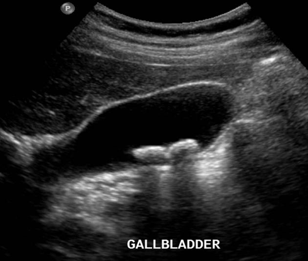
Picture 1. Abdominal ultrasound image
of the gallbladder with stones
(source: Radiopedia, CC license)

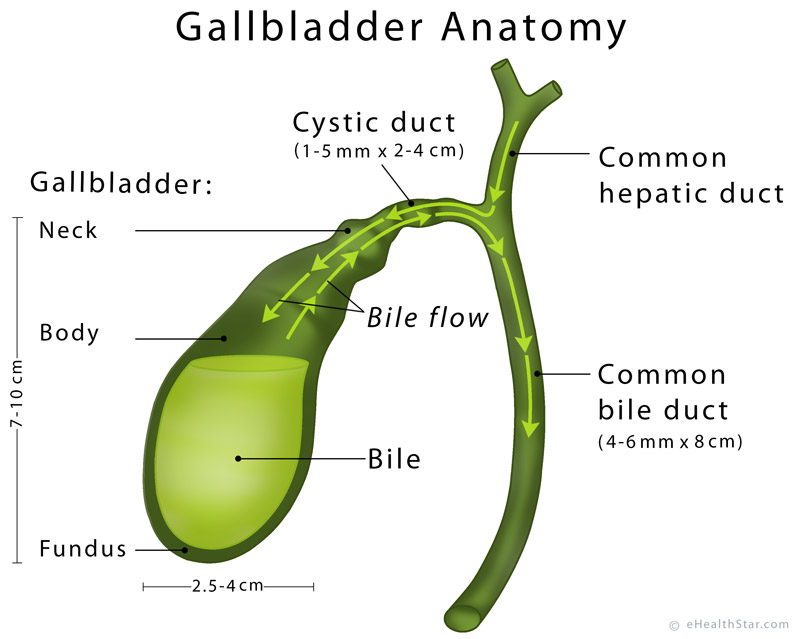
Picture 2. The gallbladder and bile ducts
Endoscopic ultrasound, which is more sensitive than standard abdominal ultrasound, involves the insertion of the probe through your mouth into the first part of the small intestine. The test can show sludge and stones as small as 0.5 mm in the gallbladder and bile ducts in more than 90% of cases [2,8,12]. It can also help in staging gallbladder cancer [7].
A HIDA scan, also called cholescintigraphy or biliary nuclear scan, involves an injection of a radioactively labeled tracer into a vein and tracking its flow in the bile through the gallbladder and bile ducts with a scanner. The test can show how well the gallbladder fills with the bile. A HIDA scan can confirm or exclude acute cholecystitis with about 90% certainty [13,14].
Another version of a HIDA scan called HIDA + CCK or gallbladder function test, which uses the hormone cholecystokinin (CCK) to stimulate the gallbladder contraction, shows not only how well the gallbladder fills but also how it empties. Reduced gallbladder emptying can occur in chronic cholecystitis [1,21] and functional gallbladder disorder called gallbladder dyskinesia [11]. The percent of bile that leaves the gallbladder after its contraction is known as an ejection fraction (EF). The normal EF range is 35-65%; when it is lower than 35%, a doctor usually suggests gallbladder removal [18].
A HIDA scan can also reveal the obstruction of the common bile duct and bile leak after gallbladder perforation or surgery [4].
Magnetic resonance imaging (MRI) can help to estimate the spread of gallbladder cancer better than standard abdominal ultrasound [3], but it is rarely used for diagnosing other gallbladder problems [7].
Magnetic resonance cholangiopancreatography (MRCP) involves an injection of a contrast dye into a vein and taking MR images to check its distribution within the bile ducts. The test can show gallstones and other abnormalities in the bile ducts better than standard abdominal ultrasound [5,6].
Endoscopic retrograde cholangiopancreatography (ERCP) involves inserting a flexible tube through your mouth into the first part of the small intestine, injecting a contrast dye into the common bile duct and taking an X-ray picture. An ERCP can reveal stones in the bile ducts in more than 90% of cases and enables their removal [12,19]. It also allows the diagnosis and treatment of sphincter of Oddi dysfunction [15].
Computed tomography (CT) can help in staging gallbladder cancer [10]. It is rarely used for diagnosing other gallbladder problems [5].
An X-ray is not a routine test for the gallbladder. When used as part of a general abdominal investigation, an X-ray can incidentally reveal stones, but only in about 20% of all cases [3]; it can also reveal a calcified or porcelain gallbladder [5].
Other Tests
Blood tests in uncomplicated gallstones are usually normal [5]. A high white blood cell count can be due to gallbladder inflammation [5]. Elevated bilirubin and liver enzymes can result from the obstruction of the common bile duct [5]. High levels of the tumor marker CA 19-9 can be due to advanced cancer of the gallbladder or bile ducts [9]. Such blood tests abnormalities can also result from diseases of other organs, so they need to be interpreted together with other tests.
Urine tests are rarely necessary for diagnosing gallbladder disease. High levels of bilirubin in the urine can increase the suspicion of the obstruction of the common bile duct brought up by other tests [20].
There is no home test for gallstones. The white lumps that appear in stool after “a gallbladder flush” are not gallstones, but pieces of fat [16].
If all your gallbladder tests are negative and you continue to have abdominal pain, you can ask a doctor about investigations of other organs, such as liver and kidney tests, upper endoscopy and colonoscopy.
- References
- Imaging of cholecystitis American Journal of Roentgenology
- Orfandis NT et al, Imaging Tests of the Liver and Gallbladder MSD Manual
- Brunetti JC, Imaging of Gallstones (Cholelithiasis) Emedicine
- HIDA scan Mayo Clinic
- Heuman DM; Gallstones (Cholelithiasis), Workup Emedicine
- Gallstones Merck Manuals Home Edition
- Gallbladder cancer Emedicine
- Rana SR et al, 2012, Role of endoscopic ultrasound in idiopathic acute pancreatitis with negative ultrasound, computed tomography, and magnetic resonance cholangiopancreatography PubMed Central
- Cancer Antigen 19-9 Lab Tests Online
- Grand D et al, 2004, CT of the Gallbladder: Spectrum of Disease American Journal of Roentgenology
- Toouli J, 2002, Biliary dyskinesia PubMed
- Stabuc B et al, 2008, Acute biliary pancreatitis: detection of common bile duct stones with endoscopic ultrasound PubMed
- J David Lane et al, Acalculous Cholecystitis Imaging Emedicine
- Gokhale SM et al, 2012, Role of cholescintigraphy in management of acute acalculous cholecystitis PubMed Central
- Bolek L, 2016, A New Approach to Identify Sphincter of Oddi Dysfunction Journal in Advances in Radiology and Medical Imaging
- Gallbladder cleanse Mayo Clinic
- Diagnosis and Management of Gallbladder Polyps PubMed Central
- Huckaby L et al, Biliary hyperkinesia: A potentially surgically correctable disorder in adolescents ScienceDirect
- Pasanen P et al, 1992, Ultrasonography, CT, and ERCP in the diagnosis of choledochal stones
PubMed - Bile duct obstruction MedlinePlus
- Jones MW et al, 2017, Gallbladder, cholecystitis, acalculous NCBI


