Porcelain gallbladder refers to calcification of the gallbladder wall. It is a type of chronic gallbladder inflammation (cholecystitis) that is considered a risk factor for gallbladder cancer.
A porcelain gallbladder does not function properly (does not fill with the bile) but rarely causes any symptoms [1]. It is usually accidentally discovered during investigations for other abdominal problems, mainly in women after 50 [1,7].
Porcelain gallbladder is rare; less than 0.1% of surgically removed gallbladders are calcified [7].
Pathology
When removed, a porcelain gallbladder looks brittle and bluish, like porcelain [6].
Other characteristics [1,6,7]:
- Shrunken gallbladder with a thick wall
- Calcification, which can be:
- Diffuse – it extends through the entire gallbladder wall (“complete intramural calcification”)
- Patchy – it involves only parts of the inner gallbladder layer (“selective mucosal calcification”)
- Presence of gallstones (in >90%)
- Cystic duct obstruction
Investigations
X-Ray
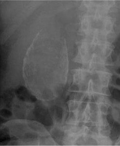
A porcelain gallbladder is visible on an X-ray image; you can see a whitish oval shadow in picture 1 below [1].
Picture 1. An x-ray image of a porcelain gallbladder
(source: Rezaie SR, Rebelem, CC license)
Computed Tomography (CT)
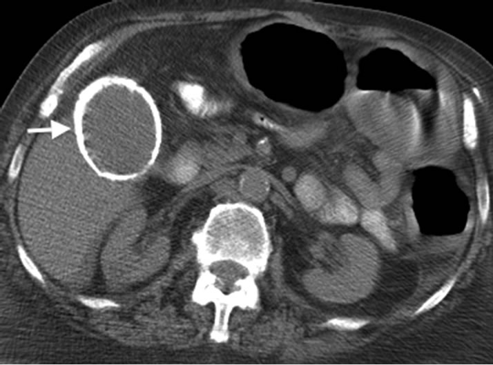
A computed tomography can provide most details about a porcelain gallbladder (Picture 2) [1].
Picture 2. A CT image of a porcelain gallbladder (arrow)
(source: Scientific Research, open access)
Ultrasound
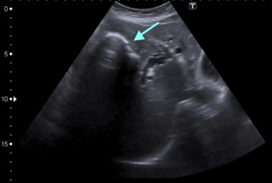
Ultrasound does not show a porcelain gallbladder very well; it can reveal calcified gallbladder wall and gallstones, though (Picture 3) [1].
Picture 3. An ultrasound image of a porcelain gallbladder
A calcified gallbladder wall (arrow) and a black “acoustic shadow” below it.
(source: Porcelain gallbladder, ScienceDirect, open access)
Porcelain Gallbladder Prognosis and Risk of Cancer
It has been long believed that 12-62% of porcelain gallbladders develops into gallbladder cancer [5], but newer studies suggest that this may occur in only about 6% [3,5,7,8]. For comparison, gallbladder cancer develops in about 1% of individuals with chronic gallbladder inflammation without calcification [3].
Patchy gallbladder calcification or”selective mucosal calcification” more likely develops into gallbladder cancer than diffuse gallbladder calcification or “complete intramural calcification” [5].
The cause-effect relationship between porcelain gallbladder and gallbladder cancer has not been clearly established [1]. Both gallbladder cancer and porcelain gallbladder share common risk factors (long-lasting chronic cholecystitis, old age), so it is not clear if porcelain gallbladder by itself is a risk factor for cancer [3,4,5,8,10,11].
Treatment
Porcelain gallbladder is usually treated by surgical gallbladder removal [9].
- References
- Khan AN, Porcelain gallbladder Emedicine
- Ashur S et al 1978, Calcified gallbladder (porcelain gallbladder) PubMed
- Schnelldorfer T, 2013, Porcelain gallbladder: a benign process or concern for malignancy? PubMed
- Radswiki et al, Porcelain gallbladder Radiopedia
- Stephen AE et al, 2001, Carcinoma in the porcelain gallbladder: a relationship revisited PubMed
- Porcelain gallbladder Learning Radiology
- Porcelain gallbladder UpToDate
- Towfigh S et al, 2001, Porcelain gallbladder is not associated with gallbladder carcinoma PubMed
- Machado NO, 2016, Porcelain gallbladder, decoding the malignant truth PubMed Central
- Chen GL et al, Porcelain Gallbladder: No Longer an Indication for Prophylactic Cholecystectomy PubMed
- Kim JH et al, 2009, Should we perform surgical management in all patients with suspected porcelain gallbladder? PubMed


